Nuclear medicine today
George de Hevesy’s work with radioactive tracers at the University of Copenhagen earned him a Nobel Prize in 1944 and was the start of an entirely new medical discipline.
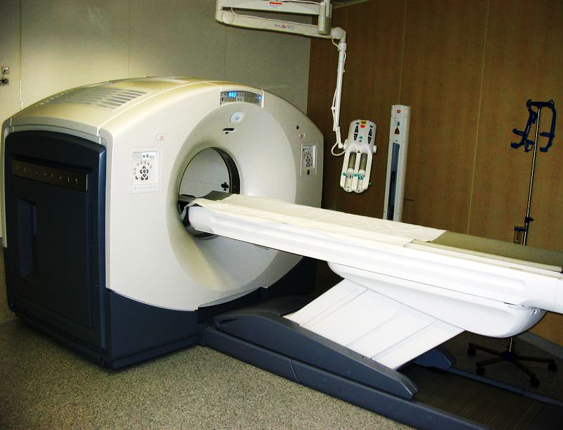
Today, nuclear medicine is used mainly in connection with PET scanning for the diagnosis of cancer, for example.
George de Hevesy’s continuing research
After George de Hevesy was awarded the Nobel Prize in 1944 and the second World War was over, he remained with his family in Stockholm, where he continued to work on the development of radioactive isotopes as tracers for nuclear medicine principles.
He worked with the development of new methods for studying metabolic diseases of the thyroid using iodine-131, the circulation of red blood cells and a host of other studies that made the development of the modern nuclear medicine scanning possible.
George de Hevesy died in 1966 at the age of 80, after which he was buried in Budapest, Hungary.
Nuclear medicine can today be described as a medical discipline that uses radioactive isotopes (radionuclides) for the diagnosis and treatment of diseases.
Nuclear medicine after Hevesy
In Denmark, nuclear medicine is mainly practiced together with clinical physiology, which became a medical specialty in 1966. Since 1982, this specialty has been designated clinical physiology and nuclear medicine.
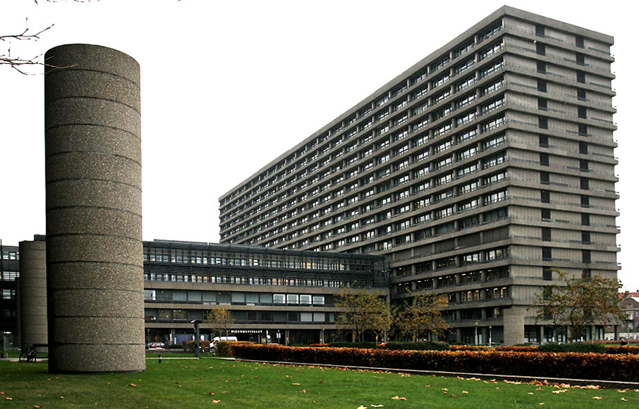
The specialty is carried out in hospital departments that are also part of the Radioactive Incident Warning System, because they have the expertise in the fields of dosimetry and radiation biology and with the necessary measuring equipment.

In the field of nuclear medicine, examinations are based on the injection of small doses of radioactive materials (a tracer), which allows the doctor to assess the function of an organ, tissue or bone.
The radioactive tracer is injected or inhaled, after which it is absorbed into the organ or area of the body to be examined.
Using a special camera or a scanner, you can take pictures and get detailed information about the material’s distribution in the body.
Many of the examinations are supplemented by CT or MR scans, which enable the researchers to simultaneously examine anatomy and function.
Nuclear medicine imaging
'Positron emission tomography' (PET) is a nuclear medicine imaging technique that employs a special type of tracers (those that disintegrate with the emission of a positron) to create three-dimensional images of body processes.
A PET scan is an imaging scan, where you inject a radioactively marked tracer into the body before scanning it. A PET scan can provide a picture of disease activity throughout the entire body and is especially used for cancer and to get a more complete picture of a disease in the entire body rather than in individual parts of the body.
The method is so sensitive that it can detect metabolic changes in the tissue at an early stage – often before the cancer can be seen on a CT or MRI scan.

Moderne scannere er kombinerede PET- og CT-scannere, der gør det muligt at optage både funktion (PET) og detaljeret anatomi (CT) under den samme undersøgelse og i den samme scanner.
Modern scanners are combined PET and CT scanners, which makes it possible to record both (PET) and detailed anatomy (CT) during the same examination and in the same scanner.
Reveals cancer and heart disease
The most common indication is for diagnosing cancer, examining whether the cancer has spread or assessing whether a treatment has worked. The most widely used tracer is FDG, a sugar, where high FDG-uptake often means a high metabolism of sugar.
Other trace elements can, for example, detect dementia or ischemic heart disease, which is characterised by a reduced oxygen supply to the heart muscle and can be expressed as angina pectoris or blood clots.
Read more about nuclear medicine:
- Website for the Department of Clinical Physiology, Nuclear Medicine and PET at Rigshospitalet in Copenhagen.
Contact

The playful researcher
“Bohr’s atomic model and quantum mechanics foundation for the development of artificial isotopes and thus the backdrop for the development of nuclear medicine. We do not think about this much in our everyday work in wards around the world, neither do think about the fact that it was Bohr’s atomic model that was the basis for our CD players, lasers, computers and so on.
The international environment at the Niels Bohr Institute in the 20s and 30s was unique, where the best gathered in a tolerant, happy, international and playful atmosphere. We can learn something from this today and it is very nice to see that the best research environments in many places around the country have a similar “homo ludens” ['playing man'] approach to research.
Basic research and support for the very best is the foundation for breakthroughs like those in the days of Bohr and Hevesy.
Only if we remember to sponsor basic research of the highest quality and provide the best research environments at universities with interdisciplinarity, an international outlook and a tolerant and open world, will there be new discoveries we do not even know we need.
When it comes to distributing research funds and designing education and research on an academic level, nationally and internationally, it is important to think about history.
The interdisciplinary research environment at Bohr’s Institute in the 30s, where science, biology and the technical sciences worked together, was incredibly fruitful and boundary-breaking research was generated at this intersection, which created both a new awareness and allowed for significant, positive changes in society, also for medicine and patient care.”
Liselotte Højgaard, Professor of Medicine and Technology at the University of Copenhagen.
