PET-scanner
When the physicist Dirac predicted the existence of antimatter in 1928, no one knew that 45 years later antimatter would be used to perform advanced brain scans.
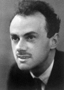
Paul Dirac predicted the existence of antimatter in 1928. Less than 50 years later scanners were built that operate using antimatter.
In 1928 physicist Paul Dirac puzzled over some mathematical equations which gave both a positive and negative result. Dirac's equations dealt with particle physics and the positive result referred to the electron. Therefore, Dirac guessed that the negative result referred to an anti-electron. This was the first time anyone suggested the existence of antimatter.
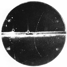
Four years later Carl D. Anderson found the particle that Dirac had written about. Anderson dubbed the particle a "positron". The positron had exactly the same mass as the electron, but is positively charged instead. Because the positron is antimatter, it cannot exist very long in our world. As soon as it runs into its antiparticle, the electron, they both disappear. At the same time they emit two gamma-photons in opposite directions.
In the 1950s American doctors got the idea that you good use these gamma-photons to produce images of the interior of the human body, which were more accurate than x-ray images.
X-ray images function by sending gamma rays through the body to a radiographic film on the other side. In this way you get an image of the structures which block the most rays - for example the bones. But if you could inject a liquid into the body, which itself emitted gamma-rays or gamma-photons, then you could photograph the movements, which the liquid makes in the body.
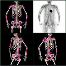
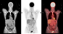
Seen here is a PET scan of how sugar water is absorbed into the muscles. The scanner produces black and white images and then they are processed and coloured in a computer programme.
3-D images of the interior of the body
Twenty years later the first PET scanner was ready. It consists of a tube, comprised of many rings, which can detect photons. And because the two gamma-photons, which are released by a positron collision, are emitted in opposite directions it is possible to determine exactly where in space they were released. Therefore, a 3D image of the places in the body the photons came from can be produced.
But in order to do this it requires of course that you have a substance that emits positrons. And there are actually some types of radioactive substances that do. Physicists can produce the specific radioactive material using a so-called cyclotron. Afterwards, chemists combine them with other substances which the body can absorb, sugar for example.
If you want the PET scanner to record where a cancerous tumour is located, you give a patient an injection of radioactive sugar water. Cancer cells absorb nutrition quicker than other cells and therefore the scanning images will show the sugar in the cancerous tumour as bright spots.
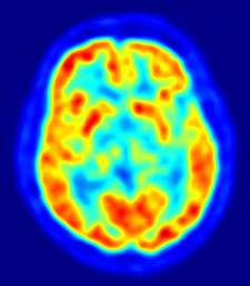
A PET scan of the brain showing the brain centres that are most active. In this image the active brain centres are red.
However, if you use a substance which remains in the bloodstream longer you can examine the blood flow in the different organs. When a brain centre is used for something it has a greater blood flow and you can therefore examine which parts of the brain are activated when the subject is asked, for example, to think of something specific or make a movement. Similarly, you can follow many other processes in the body, if only you use the correct solutions.
PET scanners are expensive because the radioactive substances only have a short lifespan so the hospital must also buy a cyclotron and employ physicists and chemists, so they can constantly produce new batchers. On the other hand, doctors can now perform advanced studies of human bodily functions which would not have been possible if physicists had not discovered the existence of that, which we call antimatter.
|
|