
Quantum Optics Colloquium
A quantum noise limited nanoparticle and biomolecule sensor & scanning nearfield imaging of optical nanofibers
This talk will have two topics: a quantum noise limited biosensor and a new method for characterisation of optical nanofibres.
Biosensors that are able to detect and track single unlabelled biomolecules are an important tool both to understand biomolecular dynamics and interactions. Recently, exceptional sensitivity has been demonstrated using the strongly enhanced fields provided by plasmonics [1,2]. However, at high field intensities photodamage to the biological specimen becomes increasingly problematic [3]. We demonstrate a quantum noise limited nanoparticle sensor capable of trapping and detecting the motion of single unlabelled biomolecules such as BSA with radius as small as 3.5 nm using 104 times lower intensity compared with state-of-the-art single molecule detectors [1,2].
Optical nanofibres are applied in a variety of new experimental platforms [4]. Especially within the fields of cold atom interfaces, bio-sensors and non-linear components a key requirement is a uniform evanescent field to ensure uniform interaction strength. Scanning electron microscopy has been the main tool for characterisation of nanofibres, but it is slow and generally damages the fibres. We demonstrate a simple and fast optical scattering-based scanning method for non-destructive characterisation of optical nanofibres. We resolve structures in the nanofibre radius with sub-nanometre precision at video rates (0.7 nm in 10 ms) producing a full image of a fibre in minutes after fabrication.
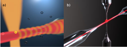
Figure 1: a) Nanoparticles/biomolecules get trapped in the evanescent field (red) around an optical nanofibre. A single trapped particle scatters light from the probe field (orange) into the guided mode of an optical nanofibre. The trapping field is 70 MHz frequency shifted from the probe field; hence the collected probe field interferes with the trapping field creating a 70 MHz heterodyne beat note signal. The signal is thus amplified and frequency shifted away from low frequency laser noise and thereby the probe field can be detected at the quantum noise limit. b) After fabrication of a nanofibre (sample fibre) it is characterised by placing a second tapered fibre (probe fibre) on to it. Light is sent through the sample fibre and transmission is measured. The probe fibre will scatter a certain fraction of the evanescent field of the sample fibre. By moving the probe fibre along the axis of the sample fibre, only the evanescent field strength changes and hence the relative field strength can be mapped out.
References
[1] Venkata R. Dantham, Stephen Holler, Curtis Barbre, David Keng, Vasily Kolchenko, and Stephen Arnold. “Label-Free Detection of Single Protein Using a Nanoplasmonic-Photonic Hybrid Microcavity ” Nano letters (2013)
[2] Yuanjie Pang and Reuven Gordon “ Optical Trapping of a Single Protein ” Nano letters (2011)
[3] K. C. Neuman, E. H. Chadd, G. F. Liou, K. Bergman, and S. M. Block, “Characterization of photodamage to Escherichia coli in optical traps,” Biophys. J. 77, 2856-2863 (1999).
[4] Limin Tong, Fei Zi, Xin Guo, Jingyi Lou "Optical microfibers and nanofibers: A tutorial" Opt. Commun. 285, 4641–4647 (2012)