BendixLab - Biophotonics & Mechanobiology

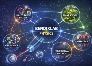
Dive into the intersection of biology and physics. Our mission is to decode how cells convert force, shape, and motion into biological function. To achieve this, we work at the crossroads of theoretical physics, molecular and cell biology, medicine, nanoscience, and plant science, creating a shared quantitative framework that enables truly interdisciplinary collaboration.

We investigate how cells use physics to organize their cell surface proteins through phase separation and geometry and how cells sense mechanical signal from their environment. Explore our findings in GPCR mechanosensing, cell surface dynamics, repair and organization using interesting new experimental methods.


We are a group of enthusiastic and creative scientists interested in understanding physical properties of biological systems at the single molecule to whole cell level.

The group has a strong interdisciplinary profile with a number of close collaborators in biology, nanoscience, medicine, chemistry and theory (see staff section for more details).

This is a short film about our research. Watch the full film here.

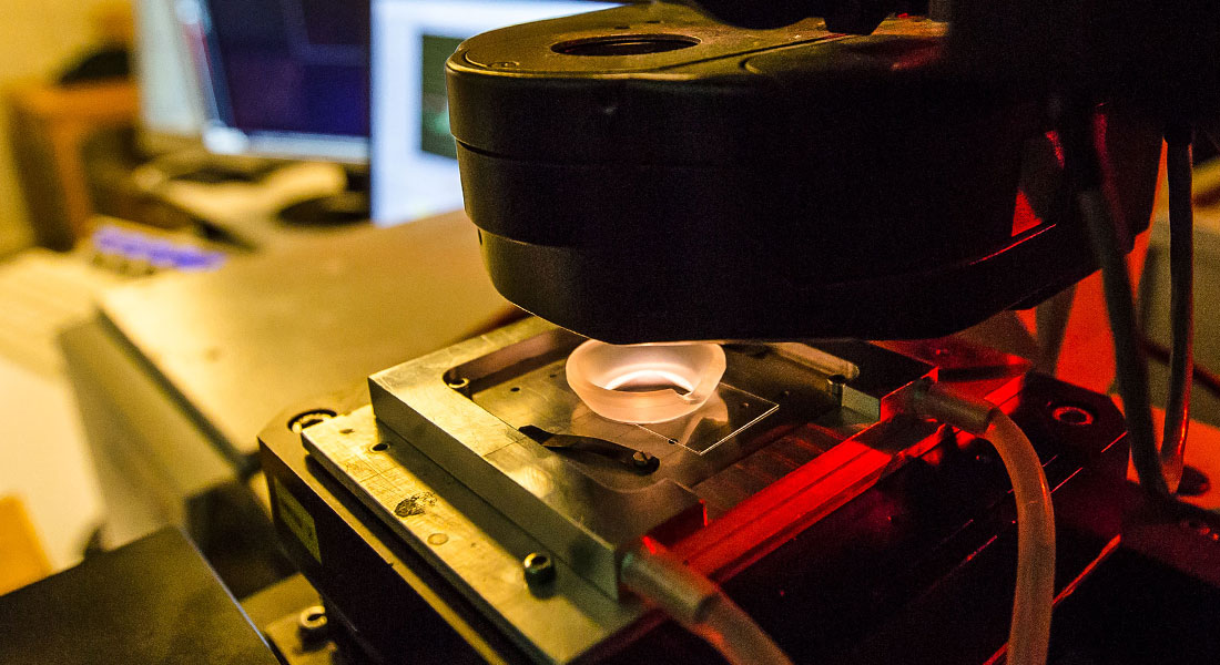
We have dual optical trapping platform combined with a confocal microscope which allows cutting edge experiments to be performed. Also, we have super resolution microscopy based on the STORM method. For imaging larger specimens, like embryos, we have light sheet microscopy which facilitates fast and low photo-toxicity imaging. Microscopic physical properties of cells are quantified using an optical trap as a force sensor/actuator whereas whole cell properties can be explored using the cell deformation cytometry.

Additionally, we have an extensive expertise in working with model membrane systems and also isolated plasma membrane systems containing the membrane proteins from the cell.

We are located at the Niels Bohr Institute in the Niels Bohr Building at the University of Copenhagen.

Funding our research

Funding the research


Research at the interface between biophotonics and mechanobiology

We explore how cells respond to physical stimuli such as membrane curvature and mechanical forces. We investigate how optical tweezers can be used to study mechanobiology, including mechanosensing and interactions at the single-molecule level. Read on to learn more about our projects and results.

Mechanosensing by GPCR

In collaboration with the Biomedical Institute at UCPH we investigate how G-protein-coupled receptors (GPCRs) (a major family of cell-surface receptors) respond to mechanical forces. This includes advanced techniques to manipulate and measure forces at the single-molecule level, providing insight into signaling mechanisms.

Our research contributes to a deeper understanding of how cells sense and respond to their mechanical environment.

Mechanosensing by GPCR

By using optical tweezers or other mechanical tools on cells we apply local or global mechanical stresses to the cells. The biochemical response is monitored using miniGs which bind to GPCRs upon activation.

This work is part of an interdisciplinary consortium called GPCRmec, funded by the Novo Nordisk Foundation, with multiple academic partners.

Membrane curvatures

Membrane curvature plays a crucial role in cellular processes.

Membrane curvatures

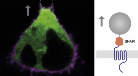
We investigate how proteins generate and sense or respond to membrane curvature, which is essential for processes such as vesicle formation, cell migration, and filopodia formation. This involves the use of advanced microscopy techniques in parallel with biophysical methods such as optical tweezers to deform membranes into high membrane curvatures.

Review: Emerging Topics in Life sci. (2023) Ruhoff et al.

Review: BioChemical Society Trans. (2023), Ruhoff et al.

Soft Matter (2021) Florentsen et al.

ACS Central Science (2020), Larsen et al.

ACS Nano (2019) Moreno-Pescador

Nature Chemical Biology (2017), Rosholm et al.

Soft Matter (2015), Barooji et al.

Sci. Rep. (2013), Ramesh et al.

Filopodia pulling and twisting dynamics

We discovered a new way that filopodia work. Filopodia are thin, finger-like protrusions that cells use as “feelers” to sense and grab their surroundings. They are found on many cell types and are especially common on moving immune cells and cancer cells where they facilitate invasion.

Filopodia

We found that filopodia can rotate because their inner support structure—made of actin fibers—can twist like a rope. This was the first clear evidence that a bundled set of actin fibers can twist in this way. We also suggested a simple physical explanation for how the twisting happens.

This twisting matters because it can help the cell pull on things it touches—similar to how a twisted rubber band tends to tighten and pull as it relaxes.

Thermoplasmonics for biology

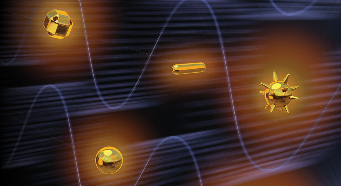
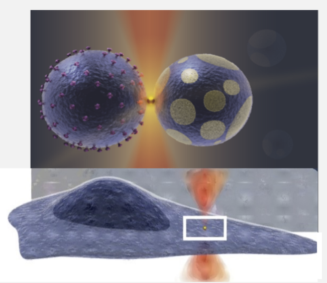
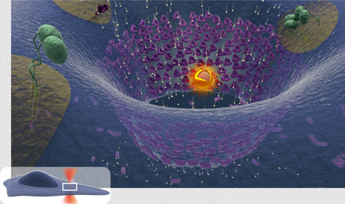
We have been pioneers in the field of plasmonics in biology. We have quantified and mapped out the optical heating from various nanostructures both theoretically and experimentally. Subsequently, we have used thermoplasmonics—tiny metal structures that can be heated with light—to do very precise manipulation or “microsurgery” on cells or soft materials.

Thermoplasmonics for biology

With this method, we can make controlled, local damage to the cell surface or even the membrane around the nucleus (in collaboration with the Cancer Institute). This lets us study how cells sense and repair injuries, which happen often when cells squeeze and move through tissue—especially immune cells and cancer cells.

We also use the same light-based heating tool for bioengineering: for example, to merge (fuse) cell membranes with synthetic membranes, or to locally “switch” membranes into a different physical state in a specific spot (a bit like changing a small patch of butter from solid to soft without affecting the rest).

Single Molecule Biophysics

Using advanced optical trapping (4 trap system from Lumicks) together with microfluidics and confocal imaging, we explore the DNA-protein interactions taking place during replication.

In collaboration with researchers from molecular biology at UCPH we investigate an array of transcription factors and proteins which help organize the genome.

Single Molecule Biophysics

Plant Biophysics

In collaboration with the Staffan Persson Lab (Plant Science, UCPH), Guillermo Pescador (UCPH) and Alexander Rohrbach (Freiburg), we have developed a new label-free microscopy approach that detects intracellular dynamics in plant cells through light scattering. Using this method, we can reveal and quantify previously hidden diffusion dynamics of intracellular vesicles in plant roots.

Plant Biophysics In collaboration with the Staffan Persson Lab (Plant Science, UCPH), Guillermo Pescador (UCPH) and Alexander Rohrbach (Freiburg), we have developed a new label-free microscopy approach that detects intracellular dynamics in plant cells through light scattering. Using this method, we can reveal and quantify previously hidden diffusion dynamics of intracellular vesicles in plant roots.  We also study the biophysics of the plant cell surface using optical trapping and thermoplasmonics as manipulation tools. In particular, we investigate how membrane phase behavior, tension and membrane curvature shape the lateral distribution of proteins.  Frontiers in Plant Science (2023). Ebrahimi et al.

We also study the biophysics of the plant cell surface using optical trapping and thermoplasmonics as manipulation tools. In particular, we investigate how membrane phase behavior, tension and membrane curvature shape the lateral distribution of proteins.

Cell membrane repair and biophysics of annexins

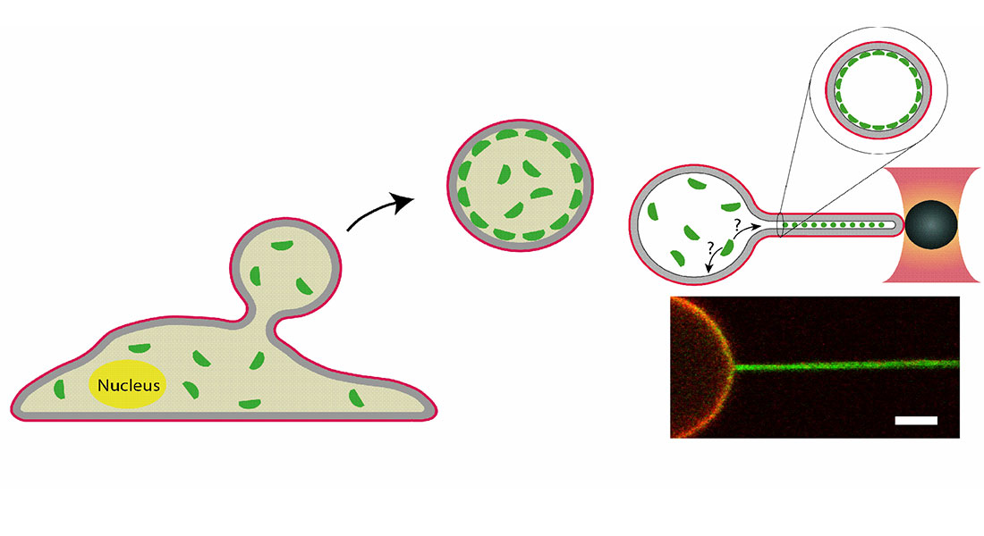
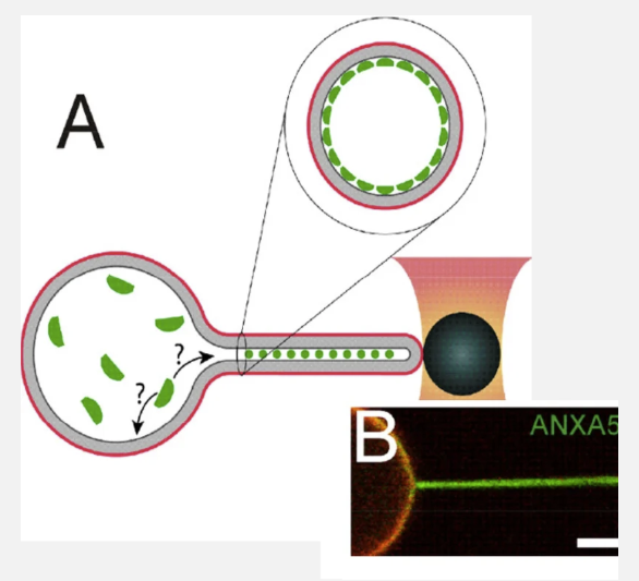
In collaboration with Jesper Nylandsted at the Danish Research Center we have investigated how cells repair surface lesions by recruiting different types of annexins. We have locally punctures living cells while monitoring recruitment of fluorescent annexins to the site of damage.

Cell membrane repair and biophysics of annexins

To gain biophysical insight into the mechanism of annexins we have made in vitro studies using membrane vesicles with encapsulated annexins. These vesicles were optically shape modulated or punctures to understand the reaction of annexins to shape and to formation  membrane holes.

Thermoplasmonics is currently being used to study the repair mechanisms of nuclear membranes as well.


Her is an overview of our main experimental facilities which our lab members and collaborators have free access to.

Optical trapping infrastructure

We offer experimental services and collaborative opportunities for researchers who want to explore optical trapping—from single-molecule biophysics to experiments with cells.

Our platform is the newest and most advanced optical trapping system currently available. It combines four independently controlled optical traps with confocal and super-resolution microscopy, integrated microfluidics, and a highly user-friendly interface—enabling a broad range of high-impact experiments with precise control and flexibility.

Optical trapping infrastructure

Fluorescent Microscopy

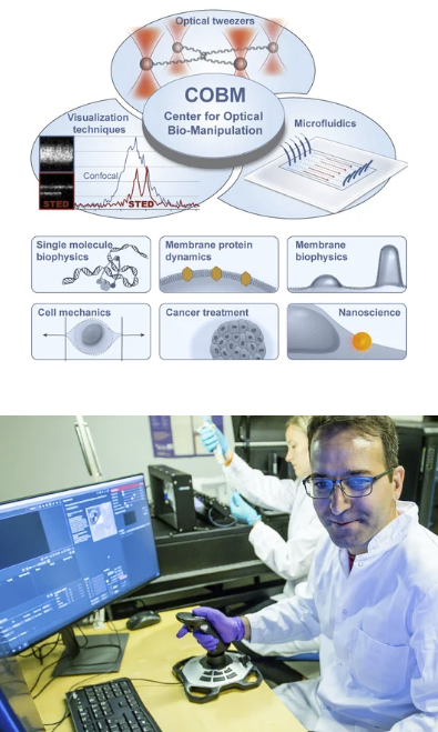
Our lab has three confocal microscopes and STORM superresolution microscopy for imaging everything from cells to single molecules. These systems are coupled to other systems such as optical tweezers or mechanical imaging (Brillouin Microscopy).

Fluorescent Microscopy

We have all typical laser wavelengths for excitation and photomultiplier tubes for collection of signals.

We offer committed collaborations of alternatively a pay-per-use basis.

Please contact us for more information.

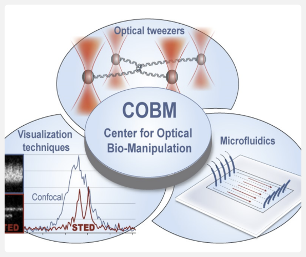
Link to infrastructure: Center for Optical Bio-Manipulation (COBM)

Brillouin Imaging

Want to image mechanical properties of matter? Then try Brillouin Microscopy which is a microscopy combining acoustics with laser scattering to achieve images showing the mechanical stiffness of your material.

Brillouin Imaging

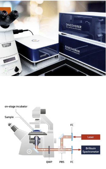
We use this for imaging living cells. This equipment is shared with Doostmohammadi Lab at the Niels Bohr Academy, UCPH

Scattering/interferometric microscopy

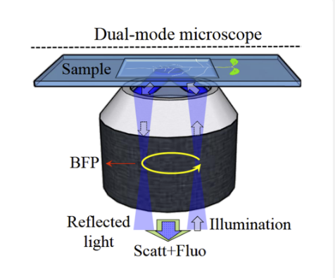
Scattering and interferometric microscopy allows us to detect nanoscopic objects with no fluorescence. This label free technology permits fast imaging at millisecond timescale or can be used for long term imaging of plant cells. Plant cells are particularly interesting for interferometric imaging due to the plant wall barrier which prevents use of intracellular markers.

Scattering/interferometric microscopy

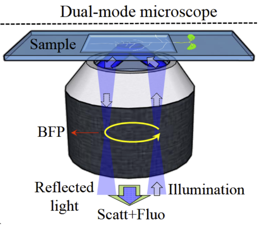
Our microscope is a Rotating Optical Coherence Scattering (ROCS) microscope which has a lateral resolution of down to 150nm and axial resolution down to 10 nm.

Mechanical perturbation

We also modulate cell shapes by mechanical action. Squeezing cells between a glass coverslip and an adjustable piston allows accurate compression of cells with 1 micrometer axial resolution. This way we can test cell response to confined conditions which are often experienced by living cells in vivo.

Mechanical perturbation

Periodic stretching of a cell substrate is an alternative mechanical method for deformation of cells. Cells can be fixed just after being stretched to detection any molecular or structural responses due to the stretching.

The cell squeezer can operate while imaging cells using confocal microscopy


We bridge other scientific fields with experimental biophysics. Our collaborators work in diverse fields such as molecular, theoretical, medical and plant science.

Interdisciplinarity

GPCRmec

This consortium includes collaborators from the medical institute and Chemistry. The goal is to understand how mechanics influences g-protein coupled receptor signaling. Financed by a Novo interdisciplinary synergy grant.

SEECLEAR

An unconventional collaboration between theoretical and experimental physics and plant science. Our goal is to understand formatiomn of the secondary cell wall. Financed by a Novo exploratory synergy grant.

Active collaborations

Signe Mathiasen

Biomedical Institute, UCPH

Staffan Persson

Plant Science, UCPH

Weria Pezheskian

Theoretical physics, Niels Bohr Institute, UCPH

Julien Duxin

Biotech Research & Innovation Centre, UCPH

Pétur Heiðarsson

Department of Biology, UCPH

Jesper Nylandsted

Danish Research Center, UCPH

Karen Martinez

Chemistry, UCPH

Mette Rosenkilde

Biomedical Institute, UCPH

Kalina Haas

INRAE, Paris, France

Jakub Sedzinski

Novo Nordisk Foundation
Center for Stem Cell Medicine, reNEW
, UCPH


1.
Ruhoff, V. T., Leijnse, N., Doostmohammadi, A. & Bendix, P. M. Filopodia: integrating cellular functions with theoretical models. Trends in Cell Biology 35, 129–140 (2025).
2.
Ruhoff, V. R. et al. BPS2025-Biophysical applications of photothermal materials. Biophysical Journal 124, 504a (2025).
3.
4.
Ruhoff, V. T., Arastoo, M. R., Moreno-Pescador, G. & Bendix, P. M. Biological applications of thermoplasmonics. Nano Letters 24, 777–789 (2024).
5.
Ruhoff, V. T. et al. Innovative thermoplasmonic fusion reveals the phase affinity of membrane proteins. Biophysical Journal 123, 68a (2024).
6.
Danielsen, H. M. D., Arastoo, M. R., Moreno-Pescador, G. & Bendix, P. M. A thermoplasmonic approach for investigating plasma membrane repair in living cells and model membranes. J vis Exp 203, e65776 (2024).
7.
Bendix, P. M. et al. Twisting tales: The emergent twist in filopodia dynamics and its role in cellular function. Biophysical Journal 123, 178a (2024).
8.
Ruhoff, V. T., Bendix, P. M. & Pezeshkian, W. Close, but not too close: a mesoscopic description of (a) symmetry and membrane shaping mechanisms. Emerging Topics in Life Sciences 7, 81–93 (2023).
9.
Moreno-Pescador, G. et al. Unlocking the secrets of membrane protein dynamics with innovative optical trapping-assisted fusion. in EUROPEAN BIOPHYSICS JOURNAL WITH BIOPHYSICS LETTERS vol. 52 S145–S145 (SPRINGER ONE NEW YORK PLAZA, SUITE 4600, NEW YORK, NY, UNITED STATES, 2023).
10.
Moreno-Pescador, G. et al. Thermoplasmonic vesicle fusion reveals membrane phase segregation of influenza spike proteins. Nano Letters 23, 3377–3384 (2023).
11.
Ebrahimi, S., Moreno-Pescador, G., Persson, S., Jauffred, L. & Bendix, P. M. Label-free optical interferometric microscopy to characterize morphodynamics in living plants. Frontiers in Plant Science 14, 1156478 (2023).
12.
Brinkenfeldt, N. K., Dias, A., Moreno-Pescador, G., Bendix, P. M. & Martinez, K. L. Quantitative Determination of Protein Concentrations in Living Cells. bioRxiv 2023–05 (2023).
13.
Bendix, P. M. et al. Active generation of twist in filopodia. in EUROPEAN BIOPHYSICS JOURNAL WITH BIOPHYSICS LETTERS vol. 52 S47–S47 (SPRINGER ONE NEW YORK PLAZA, SUITE 4600, NEW YORK, NY, UNITED STATES, 2023).
14.
Bendix, P. et al. Mechanisms of Annexin-Mediated Membrane Shaping and Curvature Sensing During Plasma Membrane Repair. in MOLECULAR BIOLOGY OF THE CELL vol. 34 48–49 (AMER SOC CELL BIOLOGY 8120 WOODMONT AVE, STE 750, BETHESDA, MD 20814-2755 USA, 2023).
15.
Ruhoff, V. T., Moreno-Pescador, G., Pezeshkian, W. & Bendix, P. M. Strength in numbers: effect of protein crowding on the shape of cell membranes. Biochemical Society Transactions 50, 1257–1267 (2022).
16.
17.
Moreno-Pescador, G. S. & Bendix, P. M. rsc. li/nanoscale. (2022).
18.
Moreno-Pescador, G. S. et al. Thermoplasmonic nano-rupture of cells reveals annexin V function in plasma membrane repair. Nanoscale 14, 7778–7787 (2022).
19.
Moreno-Pescador, G., Arastoo, M. R., Chiantia, S., Daniels, R. & Bendix, P. M. Thermoplasmonic induced vesicle fusion for investigating membrane protein phase affinity. bioRxiv 2022–09 (2022).
20.
Leijnse, N. et al. Filopodia rotate and coil by actively generating twist in their actin shaft. Nature communications 13, 1636 (2022).
21.
Heltberg, M., von Borries, M., Bendix, P. M., Oddershede, L. B. & Jensen, M. H. Temperature Controls Onset and Period of NF-κ B Oscillations and can Lead to Chaotic Dynamics. Frontiers in Cell and Developmental Biology 10, 910738 (2022).
22.
Pescador, G. S. M. et al. Investigating plasma-membrane repair employing thermoplasmonics. Biophysical Journal 120, 45a (2021).
23.
24.
Florentsen, C. D. et al. A Newly Discovered Class of Curvature Sensitive Proteins: Trimeric Annexins. Biophysical Journal 120, 285a (2021).
25.
Florentsen, C. D. et al. Annexin A4 senses membrane curvature in a density dependent manner. BiorXiv 2021–11 (2021).
26.
Christoffer Dam Florentsen, P. M. B., Alexander Kamp-Sonne, Guillermo Moreno-Pescador, Weria Pezeshkian, Ali Asghar Hakami Zanjani, Himanshu Khandelia, Jesper Nylandsted. Annexin A4 trimers are recruited by high membrane curvatures in Giant Plasma Membrane Vesicles. Soft Matter 17, 308–318 (2021).
27.
28.
Arastoo, M. R. et al. Phase Partitioning of influenza Virus Neuraminidase in a Model Membrane. Biophysical Journal 120, 48a (2021).
29.
Purohit, P., Samadi, A., Bendix, P. M., Laserna, J. J. & Oddershede, L. B. Optical trapping reveals differences in dielectric and optical properties of copper nanoparticles compared to their oxides and ferrites. Scientific Reports 10, 1198 (2020).
30.
Larsen, J. B. et al. How membrane geometry regulates protein sorting independently of mean curvature. ACS central science 6, 1159–1168 (2020).
31.
32.
Florentsen, C. D., Moreno-Pescador, G. S., Kjær, A., Oddershede, L. B. & Bendix, P. M. Plasmonic material engineering for targeted therapeutics. Advanced Optical Materials 8, 2000616 (2020).
33.
34.
Samadi, A., Jauffred, L., Klingberg, H., Bendix, P. M. & Oddershede, L. B. Optical control of strongly absorbing nanoparticles and their potential for photothermal treatment. in Complex Light and Optical Forces XIII vol. 10935 94–103 (SPIE, 2019).
35.
Moreno-Pescador, G. et al. Effect of local thermoplasmonic heating on biological membranes. in Optical trapping and optical micromanipulation XVI vol. 11083 24–34 (SPIE, 2019).
36.
37.
Moreno-Pescador, G. et al. Quantification of protein dynamics in nanotubes pulled from transfected cell-derived vesicles. in EUROPEAN BIOPHYSICS JOURNAL WITH BIOPHYSICS LETTERS vol. 48 S159–S159 (SPRINGER 233 SPRING ST, NEW YORK, NY 10013 USA, 2019).
38.
Jauffred, L., Samadi, A., Klingberg, H., Bendix, P. M. & Oddershede, L. B. Plasmonic heating of nanostructures. Chemical reviews 119, 8087–8130 (2019).
39.
Florentsen, C. D., Pescador, G. S. M., Sonne, A. K., Nylandsted, J. & Bendix, P. M. Annexin V is a Sensor of Negative Plasma Membrane Curvature. Biophysical Journal 116, 519a (2019).
40.
41.
Moreno-Pescador, G. S. et al. Lateral Distribution and Mobility of Transmembrane Proteins in Plasma Membrane Vesicles. Biophysical Journal 114, 600a (2018).
42.
Florentsen, C. D. et al. Quantification of loading and laser-assisted release of RNA from single gold nanoparticles. Langmuir 34, 14891–14898 (2018).
43.
Bahadori, A., Moreno-Pescador, G., Oddershede, L. B. & Bendix, P. M. Remotely controlled fusion of selected vesicles and living cells: a key issue review. Reports on Progress in Physics 81, 032602 (2018).
44.
Samadi, A., Bendix, P. M. & Oddershede, L. B. Optical manipulation of individual strongly absorbing platinum nanoparticles. Nanoscale 9, 18449–18455 (2017).
45.
Rosholm, K. R. et al. Membrane curvature regulates ligand-specific membrane sorting of GPCRs in living cells. Nature chemical biology 13, 724–729 (2017).
46.
Oddershede, L. B., Bahadori, A. & Bendix, P. M. Optical manipulation of hot nanoparticles can mediate selected cell fusion. in Optical Manipulation Conference vol. 10252 9–11 (SPIE, 2017).
47.
Norregaard, K. et al. Using Optically Manipulated Metallic Nanoparticles for Cancer Treatment. in Optical Trapping Applications OtTu2E-4 (Optica Publishing Group, 2017).
48.
Kamilla Norregaard, A. K., Jesper T. Jørgensen, Marina Simón, Fredrik Melander, Lotte K. Kristensen, Poul M. Bendix, Thomas L. Andresen, Lene B. Oddershede. 18F-FDG PET/CT-based early treatment response evaluation of nanoparticle-assisted photothermal cancer therapy. PloS One (2017).
49.
Bahadori, A., Oddershede, L. B. & Bendix, P. M. Optically controlled fusion of selected cells and vesicles using plasmonic nanoheaters. (2017).
50.
Bahadori, A., Oddershede, L. B. & Bendix, P. M. Hot-nanoparticle-mediated fusion of selected cells. Nano Research 1–12 (2017).
51.
Bahadori, A., Oddershede, L. & Bendix, P. Light Robotics - Structure-mediated Nanobiophotonics. in http://www.nbi.dk/∼bendix/PDFsPublications/Bahadori2017book.pdf eBook-ISBN (Elsevier, 2017).
52.
53.
54.
Barooji, Y. F., Rørvig-Lund, A., Semsey, S., Reihani, S. N. S. & Bendix, P. M. Dynamics of membrane nanotubes coated with I-BAR. Scientific reports 6, 30054 (2016).
55.
Baroji, Y. F., Rørvig-Lund, A., Reihani, S. N., Semsey, S. & Bendix, P. M. Dynamics of Membrane Tubes Formed by I-BARS. Biophysical Journal 110, 243a (2016).
56.
Bahadori, A., Lund, A. R., Semsey, S., Oddershede, L. B. & Bendix, P. M. Controlled cellular fusion using optically trapped plasmonic nano-heaters. in Optical Trapping and Optical Micromanipulation XIII vol. 9922 125–134 (SPIE, 2016).
57.
58.
Stamou, D. et al. Membrane curvature regulates the localization of G protein coupled receptors and Ras isoforms. Biophysical Journal 108, 95a–96a (2015).
59.
Rørvig-Lund, A., Bahadori, A., Semsey, S., Bendix, P. M. & Oddershede, L. B. Vesicle fusion triggered by optically heated gold nanoparticles. Nano letters 15, 4183–4188 (2015).
60.
Norregaard, K., Jørgensen, J. T., Bendix, P. M., Kjær, A. & Oddershede, L. B. Comparison of the Photothermal Efficiency of Different Types of Plasmonic Nanoparticles in vitro and in vivo. Biophysical Journal 108, 171a (2015).
61.
Leijnse, N., Oddershede, L. B. & Bendix, P. M. Helical buckling of actin inside filopodia generates traction. Proceedings of the National Academy of Sciences 112, 136–141 (2015).
62.
Leijnse, N., Oddershede, L. B. & Bendix, P. M. Helical Buckling in Filopodia. Biophysical Journal 108, 139a (2015).
63.
Leijnse, N., Oddershede, L. B. & Bendix, P. M. Dynamic buckling of actin within filopodia. Communicative & Integrative Biology 8, e1022010 (2015).
64.
Leijnse, N., Oddershede, L. B. & Bendix, P. M. An updated look at actin dynamics in filopodia. Cytoskeleton 72, 71–79 (2015).
65.
Ramesh, P. et al. Sensing and Stiffening of Tubular Membranes by the Syndapin 1 FBAR. Biophysical Journal 106, 714a (2014).
66.
Ma, H., Tian, P., Pello, J., Bendix, P. M. & Oddershede, L. B. Heat generation by irradiated complex composite nanostructures. Nano letters 14, 612–619 (2014).
67.
Barooji, Y. F., Oddershede, L., Reihani, S. N. S. & Bendix, P. M. Physical characterization of phospholipid nanotubes and the effect of BAR domain proteins on their mechanical stability. Journal, vol 43, 595–602 (2014).
68.
Baroji, Y. F., Oddershede, L. B., Seyed Reihani, S. N. & Bendix, P. M. Fluorescent quantification of size and lamellarity of membrane nanotubes. European Biophysics Journal 43, 595–602 (2014).
69.
Andersen, T., Kyrsting, A. & Bendix, P. M. Local and transient permeation events are associated with local melting of giant liposomes. Soft Matter 10, 4268–4274 (2014).
70.
Andersen, T. et al. Nanoscale phase behavior on flat and curved membranes. Nanotechnology 25, 505101 (2014).
71.
van Lengerich, B., Rawle, R. J., Bendix, P. M. & Boxer, S. G. Individual vesicle fusion events mediated by lipid-anchored DNA. Biophysical journal 105, 409–419 (2013).
72.
73.
Ott, D., Bendix, P. M. & Oddershede, L. B. Revealing hidden dynamics within living soft matter. ACS nano 7, 8333–8339 (2013).
74.
Nørregaard, K., West, A.-K. V., Bendix, P. M. & Oddershede, L. B. Nanoparticle Mediated Photothermal Therapy and Integrated miRNA Delivery. in Optical Molecular Probes, Imaging and Drug Delivery JW3B-4 (Optica Publishing Group, 2013).
75.
Kyrsting, A., Bendix, P. M. & Oddershede, L. B. Measuring the focal intensity distribution reveals nanoparticle trapping positions. in Optical Trapping Applications JT2A-32 (Optica Publishing Group, 2013).
76.
Kyrsting, A., Bendix, P. M. & Oddershede, L. B. Mapping 3D focal intensity exposes the stable trapping positions of single nanoparticles. Nano letters 13, 31–35 (2013).
77.
Bendix, P. M., Jauffred, L., Norregaard, K. & Oddershede, L. B. Optical trapping of nanoparticles and quantum dots. IEEE journal of selected topics in quantum electronics 20, 15–26 (2013).
78.
Rawle, R. J., van Lengerich, B., Martin Bendix, P., Chung, M. & Boxer, S. G. DNA-Mediated Fusion Between Small Vesicles and a Planar, Tethered Bilayer Patch. Biophysical Journal 102, 605 (2012).
79.
Ma, H., Bendix, P. M. & Oddershede, L. B. Measurements of extreme orientation-dependent temperature increase around an irradiated gold nanorod. in Optical Trapping and Optical Micromanipulation IX vol. 8458 246–255 (SPIE, 2012).
80.
Ma, H., Bendix, P. M. & Oddershede, L. B. Large-scale orientation dependent heating from a single irradiated gold nanorod. Nano letters 12, 3954–3960 (2012).
81.
Kyrsting, A., Bendix, P. M. & Oddershede, L. B. Photothermal heating of optically trapped gold nanoparticles quantified using controlled vesicle cargo release. in Optical Trapping and Optical Micromanipulation IX vol. 8458 132–139 (SPIE, 2012).
82.
Bendix, P. M. & Oddershede, L. Optical Manipulation of Nano-Scale Vesicles. Biophysical Journal 102, 87a (2012).
83.
van Lengerich, B., Rawle, B. J., Bendix, P. M., Chung, M. & Boxer, S. G. Individual Vesicle-Vesicle and Vesicle-Planar Bilayer Fusion Events Mediated by DNA. Biophysical Journal 100, 633a (2011).
84.
van Lengerich, B., Bendix, P. M. & Boxer, S. G. DNA-Mediated Fusion Between Small Vesicles and a Planar, Tethered Bilayer Patch. Biophysical Journal 101, L37–L39 (2011).
85.
Stamou, D. G., Bendix, P. M., Wibroe, P. P. & Hatzakis, N. S. Static and Dynamic Disorder Observed in the Phase Transition Behavior of Individual Small Unilamellar Vesicles. Biophysical Journal 100, 35a (2011).
86.
Rawle, R. J., van Lengerich, B., Chung, M., Bendix, P. M. & Boxer, S. G. Vesicle fusion observed by content transfer across a tethered lipid bilayer. Biophysical Journal 101, L37–L39 (2011).
87.
Kyrsting, A., Bendix, P. M., Stamou, D. G. & Oddershede, L. B. Heat profiling of three-dimensionally optically trapped gold nanoparticles using vesicle cargo release. Nano letters 11, 888–892 (2011).
88.
Chung, M., Bendix, P. M., Kim, N. & Boxer, S. G. DNA-Machinery for Delivering Membrane Proteins into Free Standing Lipid Bilayers. Biophysical Journal 100, 633a (2011).
89.
Bendix, P. M. & Oddershede, L. B. Expanding the optical trapping range of lipid vesicles to the nanoscale. Nano letters 11, 5431–5437 (2011).
90.
Bendix, P., Kyrsting, A., Reihani, N. & Oddershede, L. Heating in Optically Trapped Gold Nanoparticles Measured in Artificial Membranes. in Optical Trapping Applications OTMA3 (Optica Publishing Group, 2011).
91.
Bendix, P. M., Reihani, S. N. S. & Oddershede, L. Direct Measurement of Heating by Optically Trapped Gold Nanoparticles Using Molecular Sorting in a Lipid Bilayer. Biophysical Journal 98, 185a (2010).
92.
Bendix, P. M., Reihani, S. N. S. & Oddershede, L. B. Direct measurements of heating by electromagnetically trapped gold nanoparticles on supported lipid bilayers. ACS nano 4, 2256–2262 (2010).
93.
Koenderink, G. H. et al. An active biopolymer network controlled by molecular motors. Proceedings of the National Academy of Sciences 106, 15192–15197 (2009).
94.
Bendix, P. M., Pedersen, M. S. & Stamou, D. Quantification of nano-scale intermembrane contact areas by using fluorescence resonance energy transfer. Proceedings of the National Academy of Sciences 106, 12341–12346 (2009).
95.
Bendix, P. M., Wibroe, P. P. & Stamou, D. Phase Transitions in Single Nano-Vesicles. Biophysical Journal 96, 148a (2009).
96.
Bosanac, L., Aabo, T., Bendix, P. M. & Oddershede, L. B. Efficient optical trapping and visualization of silver nanoparticles. Nano letters 8, 1486–1491 (2008).
97.
Bendix, P. M. et al. A quantitative analysis of contractility in active cytoskeletal protein networks. Biophysical journal 94, 3126–3136 (2008).
98.
Fischer, W. Sprechen Sie G-DRG. (2007).
99.
Hansen, P. M., Tolić-Nørrelykke, I. M., Flyvbjerg, H. & Berg-Sørensen, K. tweezercalib 2.0: Faster version of MatLab package for precise calibration of optical tweezers. Computer physics communications 174, 518–520 (2006).
100.
Hansen, P. M., Tolic-Nørrelykke, I. M., Flyvbjerg, H. & Berg-Sørensen, K. Catalogue identifier: ADTV_v2_1 Distribution format: tar. gz. Journal reference: Comput. Phys. Commun 174, 518 (2006).
101.
Hansen, P. M. Cellular mechanics studied by novel nano-tools and reconstituted model system. (University of Copenhagen, Niels Bohr Institute, 2006).
102.
Bendix, P. M., Tolic-Nørrelykke, I. M., Flyvbjerg, H. K. & Berg-Sørensen, K. Tweezercalib 2.1. Computer Physics Communications (2006).
103.
Hansen, P. M. & Oddershede, L. B. Optical trapping inside living organisms. in Optical Trapping and Optical Micromanipulation II vol. 5930 593003 (SPIE, 2005).
104.
Hansen, P. M., Dreyer, J. K., Ferkinghoff-Borg, J. & Oddershede, L. Novel optical and statistical methods reveal colloid–wall interactions inconsistent with DLVO and Lifshitz theories. Journal of colloid and interface science 287, 561–571 (2005).
105.
Hansen, P. M., Bhatia, V. K., Harrit, N. & Oddershede, L. Expanding the optical trapping range of gold nanoparticles. Nano letters 5, 1937–1942 (2005).
106.
Dreyer, J. K., Hansen, P. M. & Oddershede, L. B. Optical probing of specific and nonspecific interactions with nanometer resolution. in Optical Trapping and Optical Micromanipulation vol. 5514 402–409 (SPIE, 2004).
107.
108.
109.
Ramesh, P. et al. Dependent Manner.
110.
111.
Leijnse, N., Oddershede, L. B. & Bendix, P. M. Helical Buckling of Actin within Filopodia Generates Traction.
112.
113.
114.
Jørgensen, J. T. et al. Supplementary Information to.
115.
116.
117.
Andersen, T., Kyrsting, A. & Bendix, P. M. This journal is\copyright The Royal Society of Chemistry 2014.


We have many different projects for Master's students and also occasional projects for undergraduate students.

This is a video of Victoria Ruhoff explaining her project about biophysics of virus proteins (Danish).

Cellular repair system investigated by laser based nano-surgery

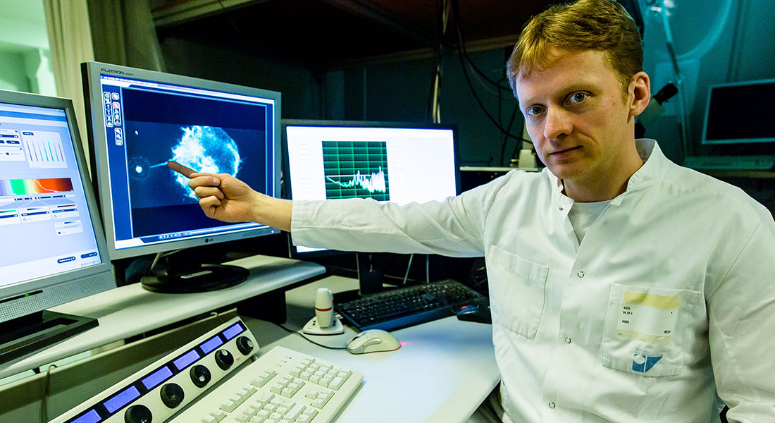
When cells migrate in the body they often experience membrane ruptures which results in excessive calcium flowing into the cell. This can be lethal unless the membrane is sealed within milliseconds. Cells have a built-in surface repair kit which is activated by calcium influx and hence can allow the cell to self-heal within seconds after injury. Cancer cells are extremely efficient in repairing their surface since they have an increased expression of various annexin proteins which are thought to be the major proteins involved in the membrane repair.

The project will involve testing the membrane repair system in cells by using thermoplasmonics to inflict a nanoscopic hole in the membrane. This will be done by irradiating a plasmonic gold nanostructure placed on the surface of the cell. Confocal microscopy and super-resolution microscopy will be used to monitor the recruitment of various annexins which are labeled with fluorescent proteins. The overall aims are to i) investigate whether invasive cancer cells are more efficient in dealing with thermoplasmonic ruptures than non-invasive cells ii) investigate the role of several different annexins in the repair process including other proteins like ESCRT and actin.

The project is a collaboration with Kræftens Bekæmpelse samt Syddansk Universitet. 

Contact: Poul Martin Bendix, bendix@nbi.dk


Patterning in large bacterial communities

In nature, bacteria actively search for a surface to form larger communities, i.e., biofilm, with extended cooperativity and defense. We know that sectors with low genetic diversity form within the colony, even among cells of similar fitness. This self-organization of microbial cell communities is the result of genetic drift in complex interplay with evolution, competition, and cooperation.

We offer various projects to explore pattern formation by growing bacteria both in vivo and in silico. We believe the close interplay between theory and experiments will provide a more complete understanding of cooperation and competition among cells in larger communities.  We aim to point out general features of growth pattern, which can be generalized in wider class of systems. In the long term, we may draw parallels to mammalian cell systems, where patterning is crucial for example in embryonic development.

Possible subprojects include:

Colony shape

The relation between the individual cell shape and the colony shape

This project combines theory and experiments depending on your interests. Experimentally, the project can include bacterial cell culture, colony growth, and advanced fluorescence microscopy. Theoretically, we plan to first simulate an individual cell-based model where the particles grow, divide, and interact through mechanical force. Depending on the development of the project, simplified lattice models or partial differential equation-based models can also be used.

Supervisors: Liselotte Jauffred & Namiko Mitarai


Signe Mathiasen

Biomedical Institute, UCPH

Staffan Persson

Plant Science, UCPH

Weria Pezheskian

Theoretical physics, Niels Bohr Institute, UCPH

Julien Duxin

Biotech Research & Innovation Centre, UCPH

Pétur Heiðarsson

Department of Biology, UCPH

Jesper Nylandsted

Danish Research Center, UCPH

Karen Martinez

Chemistry, UCPH

Mette Rosenkilde

Biomedical Institute, UCPH

Kalina Haas

INRAE, Paris, France

Jakub Sedzinski

Novo Nordisk Foundation
Center for Stem Cell Medicine, reNEW
, UCPH


Poul Martin Bendix, Group leader
E-mail: bendix@nbi.ku.dk
Tel: +45 35325251 or +45 61602454
Niels Bohr Building, Jagtvej 132
2200 Copenhagen N.

Content not available due to cookie preferences

You cannot see the content of this field because of your cookie preferences.

Click here to change your cookie settings.

Category: Marketing


Staff

Name Title Job responsibilities
Search in Name Search in Title Search in Job responsibilities
Bendix, Pól Martin Associate Professor Associate Professor, Group Leader Billede af Bendix, Pól Martin
Farhangi Barooji, Younes Academic Staff Billede af Farhangi Barooji, Younes
Hamel Ascanio, Luis Eduardo PhD Student Billede af Hamel Ascanio, Luis Eduardo
Mesi, Elsa PhD Fellow Billede af Mesi, Elsa
Moreno Pescador, Guillermo Sergio Guest Researcher Billede af Moreno Pescador, Guillermo Sergio

Master/undergraduate students

Navn Titel E-mail Foto
Natálie Palková INTERACT PhD student from Duxin Lab natalie.palkova
@bric.ku.dk
Natalie Palkova
Ciara Dooley Master Student rhp186@alumni.ku.dk
Noëlle Klasner Master Student noelle.klasner@gmail.com
Laura Isabella Pultz Henriksen Bachelor Student laura.henriksen@nbi.ku.dk
Konrad Skovmand Olsen Voluntary student kol@adm.ku.dk